Health, Fitness,Dite plan, health tips,athletic club,crunch fitness,fitness studio,lose weight,fitness world,mens health,aerobic,personal trainer,lifetime fitness,nutrition,workout,fitness first,weight loss,how to lose weight,exercise,24 hour fitness,

10/21/21

This article was previously published February 5, 2021, and has been updated with new information.

If SARS-CoV-2 has frazzled your nerves, I have bad news for you. Scientists are already cooking up more virulent and lethal versions. In a January 22, 2021, Twitter post, biotech entrepreneur Yuri Deigin highlighted a study posted on the preprint server bioRxiv at the end of December 2020, saying:1

"Ok, the prize for the craziest and most dangerous gain-of-function research goes out to Italian virologists who took SARS[-CoV-]2 and passaged it in vitro in the presence of neutralizing antibodies.2 It quickly obliged and mutated to escape them. Yay for a novel, more dangerous SARS3!"

"Passaging" refers to a genetic engineering technique where a virus is grown in a series of different animal tissue cultures. With each "pass," the virus will mutate slightly, gaining different functions.

Serial Passaging Allows Virus to Jump Species

As just one example, a potential outcome of this somewhat crude technique (considering the genetic engineering technology now available) would be that the virus could gain the ability to infect a host species it could not infect before. Some experts have speculated that this might be one way in which SARS-CoV-2 was created.

In an in-depth article3 published in New York magazine January 4, 2021, Nicholson Baker reviewed the history of viral gain-of-function research, providing the following example of serial passaging:

"Baric … described in this early paper how his lab was able to train a coronavirus, MHV, which causes hepatitis in mice, to jump species, so that it could reliably infect BHK (baby-hamster kidney) cell cultures.

They did it using serial passaging: repeatedly dosing a mixed solution of mouse cells and hamster cells with mouse-hepatitis virus, while each time decreasing the number of mouse cells and upping the concentration of hamster cells.

At first, predictably, the mouse-hepatitis virus couldn't do much with the hamster cells, which were left almost free of infection, floating in their world of fetal-calf serum.

But by the end of the experiment, after dozens of passages through cell cultures, the virus had mutated: It had mastered the trick of parasitizing an unfamiliar rodent. A scourge of mice was transformed into a scourge of hamsters …"

Scientists Have Created Coronavirus That Escapes Antibodies

So, what exactly have they come up with now? As summarized by Deigin, researchers serial passaged live SARS-CoV-2 in plasma obtained from a recovered COVID-19 patient that had a high amount of neutralizing antibodies in it.4

For clarification, you have two types of antibodies. Neutralizing antibodies are, as the name implies, antibodies that neutralize (kill) viruses and prevent infection, whereas binding antibodies cannot prevent infection.

The neutralizing antibodies in the plasma successfully and completely neutralized the virus during the first seven passages, but then, the virus mutated to evade the antibodies. As explained by the authors:5

"The plasma fully neutralized the virus for 7 passages, but after 45 days, the deletion of F140 in the spike N-terminal domain (NTD) N3 loop led to partial breakthrough. At day 73, an E484K substitution in the receptor-binding domain (RBD) occurred, followed at day 80 by an insertion in the NTD N5 loop containing a new glycan sequon, which generated a variant completely resistant to plasma neutralization."

In other words, they created a SARS-CoV-2 variant that bypasses acquired immunity and negates the immunity you normally would have after recovering from the infection. As such, it could be extremely lethal.

"Computational modeling predicts that the deletion and insertion in loops N3 and N5 prevent binding of neutralizing antibodies," the authors say, adding:

"The recent emergence in the United Kingdom and South Africa of natural variants with similar changes suggests that SARS-CoV-2 has the potential to escape an effective immune response and that vaccines and antibodies able to control emerging variants should be developed."

Selective Pressure of Vaccination May Pose a Problem

Now, further down in the paper, they point out that the reason they did this study was to determine "whether the authentic virus, under the selective pressure of the polyclonal immune response in convalescent or vaccinated people, can evolve to escape herd immunity and antibody treatment."

Since the virus can mutate to evade neutralizing antibodies, then it could potentially mutate under the "selective pressure" of vaccination as well, which in turn raises the question: If we mass vaccinate, will we end up with a more lethal virus?

The solution these researchers seem to propose is to start thinking about vaccinating people for emerging SARS-CoV-2 variants, meaning we may need to develop a new vaccine — much like the seasonal flu vaccine, — to match the circulating strains of each season.

Considering the first COVID-19 mRNA vaccines (which are most accurately described as gene therapy) are wreaking absolute havoc on people's health already, the idea of implementing a twice-a-year gene-therapy regimen against COVID-19 strikes me as assured destruction of the human race.

Is SARS-CoV-2 Result of Gain-of-Function Research in Wuhan?

Jamie Metzl is a geopolitics expert, World Health Organization adviser and senior fellow at the Atlantic Council. January 4, 2021, CBS News interviewed her about the "conspiracy theory" that SARS-CoV-2 was created in a biosecurity level 4 laboratory in Wuhan, China. Metzl believes the COVID-19 pandemic is the result of an accidental leak from that lab.

This, he says, is a logical conclusion based on the facts before us. First, Wuhan is far from the southern part of China where horseshoe bats (the supposed source host) exist.

Second, the Wuhan Institute of Virology (WIV) was known to have performed controversial gain-of-function research on bat coronaviruses and, according to U.S. diplomats who had visited the lab in 2018, significant safety shortcomings were apparent.6

Third, SARS-CoV-2's closest relative (RaTG13) has been traced back to samples collected in 2012 from miners sickened after working in an abandoned mine in Mojiang. There's no trace of the virus anywhere between 2012 and 2019, until it suddenly caused an outbreak in Wuhan.

Lastly, "We see this massive Chinese cover-up," Metzl says, "destroying samples, shutting off access to databases, imprisoning journalists [and] silencing scientists."

On top of that, Metzl points out that scientists working at the WIV have been unable to account for all the viruses in their database, and level 4 biosecurity laboratories around the world have experienced many safety breaches in the past.

Investigative Committees Are Severely Compromised

As noted by Metzl — who also published an op-ed about this in Newsweek — what we need is a full, independent, all-access forensic investigation into the origin of this virus. If we don't, we will not be ready for whatever else that might be right around the corner.

He also warned that while the WHO had assembled a committee7 to investigate, China was granted veto power to decide who would be on that committee, and the primary investigation was to be carried out by Chinese representatives. The WHO's committee would then simply review their findings. This questionable setup made it highly unlikely that we would get to the truth.

Indeed, almost immediately, and as soon as the report was made public when the WHO's committee was done with their "investigation," the members of the committee raised serious concerns about its ability to conduct an unbiased investigation. One of its members was singled out as particularly ethically compromised: Peter Daszak, Ph.D., is the president of EcoHealth Alliance, a nonprofit organization that has worked closely with the WIV.

When SARS-CoV-2 first emerged in Wuhan, the EcoHealth Alliance was actually providing funding to the WIV to collect and study novel bat coronaviruses. He has publicly and repeatedly dismissed the possibility of the pandemic being the result of a lab leak.8

However, a pile of evidence collected in the months following put huge doubts on Daszak's claims,9 so much so that a U.S. GOP House Foreign Affairs Committee member called for Daszak to be subpoenaed to testify about the"disinformation campaign designed to suppress public discussion about a potential lab leak."10

Daszak Was the Fox Guarding the Hen House

Importantly, correspondence obtained by U.S. Right to Know (USRTK) show Daszak played a central role in the plot to obscure the lab origin of SARS-CoV-2 from the very beginning by crafting a scientific statement condemning such inquiries as "conspiracy theory."11,12

This manufactured "consensus" was then relied on by the media to counter anyone presenting theories and evidence to the contrary. Daszak also was the head of a second commission to investigate the origin of the virus, The Lancet COVID-19 commission,13 thereby ensuring that the "consensus" would be maintained.

Ironically, in 2015, Daszak actually warned a global pandemic might occur from a laboratory incident and that "the risks were greater with the sort of virus manipulation research being carried out in Wuhan."14 Earlier that year, he was also a key speaker at a National Academies of Science seminar on reducing risk from emerging infectious diseases.

Among the material Daszak presented at that meeting was a paper titled, "Assessing Coronavirus Threats," which included an examination of the "spillover potential" from "genetic and experimental studies" on viruses. In particular, he highlighted the danger of experimenting on "humanized mice," meaning lab mice that have been genetically altered to carry human genes, cells or tissues.

Considering Daszak's personal involvement with gain-of-function research in general, and research efforts at WIV in particular, he had more than enough motivation to make sure the blame for the COVID-19 pandemic was not laid at the feet of researchers such as himself, especially those at WIV.

As part of these investigative committees, any conclusions they came up with was suspect. In fact, according to reports, the original WHO commission had no intention of investigating either the WIV15 or the lab escape theory!16  Not surprisingly, in June 2021, the Daily Mail reported that Daszak was removed from the COVID commission looking at the origins of the pandemic “after helping secretly denounce the lab leak theory while failing to mention his close ties to the same facility.”17

WHO Appoints Second Investigative Committee on COVID Origin

With mounting evidence that the virus may have come from a lab, whether leaked or intentional, WHO's director general Tedros Ghebreyesus has decided that a second investigation is needed. "Despite the WHO's initial findings, Tedros has called for audits of Wuhan laboratories, including the Wuhan Institute of Virology, which some scientists believe may be the source of the virus that caused the first infections in China," NPR reported.18

Ghebreyesus announced the appointment of the new, 26-member committee October 13, 2021 — and Daszak is not a part of it. In an editorial in the journal Science, Ghebreyesus wrote:19

"The newly established World Health Organization (WHO) Scientific Advisory Group on the Origins of Novel Pathogens (SAGO)20 presents an unprecedented opportunity to better guide studies that specifically investigate high-threat pathogens.

Its mandate is to advise the WHO on developing a framework to define comprehensive studies on the origins of such pathogens, including SARS-CoV-2 — information that is essential for developing policies and enhancing preparedness to reduce the possibility of future zoonotic spillover events (transmission of a pathogen from animals to humans) and the chance that those events become major outbreaks …

… it's clear that the scientific processes have been hurt by politicization, which is why the global scientific community must redouble efforts to drive the scientific process forward. In forming SAGO, experts were selected (from an open call for applicants) with diverse technical expertise from countries in all six WHO regions …

… laboratory hypotheses must be examined carefully, with a focus on labs in the location where the first reports of human infections emerged in Wuhan. A lab accident cannot be ruled out until there is sufficient evidence to do so and those results are openly shared."

Links to US Commissioned Research
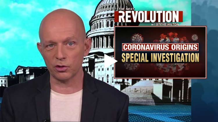
coronavirus origins special investigations
Video may not work on all browsers

>>>>> Click Here <<<<<

While most of the focus has been on the WIV, the U.S. and other Western nations are not without blame. In the video above, "The Next Revolution" host Steve Hilton reviews the origin of COVID-19, linking the outbreak to research around the world.

He starts reviewing research done by the Erasmus Centre in the Netherlands 10 years ago. There, they were able to get an influenza A/H5N1 virus to mutate and become airborne by injecting it into ferrets. This led to an explosion of gain-of-function virus research all around the world. Interestingly, that Dutch study was funded by none other than Dr. Anthony Fauci's National Institute of Allergy and Infectious Diseases (NIAID).

While the original intent may have been noble — stay a step ahead of nature so we're not surprised by natural mutations that might threaten the human population — by creating more virulent pathogens, the work itself ends up posing significant risk.

This was why, in 2014, the Obama administration put a moratorium on gain-of-function research after recent biosafety incidents had highlighted the risky nature of such study. The moratorium included pausing gain-of-function research on influenza, MERS and SARS viruses.

However, as noted by Hilton, Fauci has long been a steadfast advocate of this kind of research, and shortly before the moratorium was put into place, he had funded a project to assess the risk of bat coronavirus emergence and the "spillover potential at high-risk human-wildlife interfaces in China." At the end of that project description, they state:

"Predictive models of host range (i.e. emergence potential) will be tested experimentally using reverse genetics, pseudovirus and receptor binding essays, and virus infection experiments across a range of cell cultures from different species and humanized mice."

This is precisely the kind of research the Obama administration placed a moratorium on, but Fauci didn't drop it. Instead, he contracted it out to the EcoHealth Alliance — the group run by Daszak. Daszak himself was the project leader. Over the next six years, EcoHealth Alliance received $3.75 million for projects relating to this investigation.

Fauci, Daszak and the WIV Appear To Be Key Culprits

Daszak, in turn, subcontracted out a key piece of the research — the gain-of-function part — to the WIV. In his report, Hilton reviews some of the papers published throughout this project, proving they were indeed part of the research Fauci funded.

He points out that while many admit the NIAID funded the WIV in general, a paper co-written by Daszak and Shi Zhengli, proves Fauci funded gain-of-function research on bat coronavirus specifically.

After Hilton's team reached out to the NIH and Fauci for comment, the paper mysteriously disappeared. The paper in question, published in 2017, shows they built various chimeras based on bat coronaviruses collected. They then infected human cells with these chimeras in the lab, proving that their manmade viruses could replicate.

The genetic changes they made to these chimeras "unlocked a specific doorway to the human body," Hilton explains, and this doorway is precisely the one SARS-CoV-2 uses, namely the ACE2 receptor.

While none of the genetically engineered viruses described in that 2017 paper is identical to SARS-CoV-2, the paper proves it's possible to create these kinds of viruses using current technologies. What's more, that project continued for another three years, which puts us into 2020. During those three years, any number of new variants may have been created.

In light of the evidence, Fauci's role as chief medical adviser to the White House and leader of the coronavirus task force is "completely untenable," Hilton says. Indeed, his conflicts of interest make Fauci just as unsuitable for these roles as Daszak is for the ones to which he was assigned.

They're both involved up to their eyeballs in the research that may be the very source of this pandemic, yet both have been placed in key roles to inform, guide and direct the public on these matters. It's scientific corruption at its finest.

Surely, there are other experts out there who would be just as, if not more, qualified for these roles. "Fauci must step aside until we get to the bottom of his role in creating — unintentionally, of course — this catastrophic global pandemic," Hilton says. We also need to know whether the U.S. government is still funding research that could lead to another, even more devastating pandemic.


from Articles https://ift.tt/3jsrU7n
via IFTTT

Your colon, which is also known as the large intestine, plays an incredibly important role in your health. As part of the digestive tract, bacteria in the colon are responsible for the final breakdown of food material before it passes into the rectum and is excreted through the anus.1

New evidence published in Frontiers in Pharmacology show the antiparasitic medication ivermectin may have a new application in the treatment of colorectal cancer (CRC).2 Researchers are hopeful this may have a positive impact on colon cancer deaths. Colon cancer is the third leading cancer diagnosis and third cause of cancer death in the U.S.3

According to data from the National Cancer Institute,4 an estimated 149,500 new cases of colorectal cancer will be diagnosed in 2021 and an estimated 52,980 people will die. This represents 7.9% of all new cases of cancer diagnosed in 2021 and 8.7% of all cancer deaths.

There are modifiable risk factors associated with colorectal cancer.5 For example, lifestyle factors over which you have control that reduce your risk of colorectal cancer include your diet, alcohol consumption, activity level, weight and history of smoking.

In 2015, the International Agency for Research on Cancer, an arm of the World Health Organization,6 concluded that processed meat could cause colorectal cancer in humans and classified it as a Group 1 carcinogen. According to the WHO, this means:

“... there is convincing evidence that the agent causes cancer. In the case of processed meat, this classification is based on sufficient evidence from epidemiological studies that eating processed meat causes colorectal cancer.”

Ivermectin Shows Promise in Treatment of Colorectal Cancer

Wrongly vilified as a “livestock drug” by the media in the treatment of COVID-19 with “scant evidence it works,”7 researchers have found a new use for this Nobel Prize-winning medication.8 As the research team wrote in the published study, although CRC is the third most common cancer worldwide, it still lacks effective therapy.9

Past research has demonstrated that ivermectin also has anti-inflammatory, antitumor and antiviral properties. To test the influence ivermectin may have on colorectal cancer cells, the team used cancer cell lines SW48010 and SW1116.11 Both are epithelial cell lines from the large intestine in humans.

The researchers12 used multiple tests to determine cell viability and apoptosis after exposure to ivermectin. They also measured reactive oxygen species levels and cell cycle. To explore the effect on proliferation, the researchers used different concentrations of ivermectin on the cultured cells and found cell viability decreased in a dose-dependent and time-dependent manner.

The ivermectin also altered cell morphology, demonstrating a decrease in cells after just 24 hours and a loss of their original shape. Cultured cells were also exposed to concentrations of ivermectin after which cell viability and apoptosis were measured. The researchers found an increase in apoptosis indicating a dose-dependent effect.

Additionally, the researchers measured the activity of Caspase-3 that plays a vital role initiating apoptosis. They found that ivermectin increases Caspase 3/7 activity in both cell lines in a dose-dependent manner.

This information supports past studies that have suggested ivermectin has anticancer activity against cancers of the digestive system, reproductive system, brain, respiratory system, hematological and breast. The researchers concluded the data demonstrated:13

“... ivermectin may regulate the expression of crucial molecules … Therefore, current results indicate that Ivermectin might be a new potential anticancer drug for treating human colorectal cancer and other cancers.”

Current Colorectal Tumor Treatments Are Invasive and Damaging

The potential use of ivermectin in the treatment of colorectal cancer, or other cancers, offers great hope since current treatments are often invasive and damaging. Ivermectin has been prescribed successfully in humans for 40 years14 with a known side effect profile. This includes drowsiness, headache, mild skin rash, nausea, diarrhea and dizziness.15

The American Cancer Society’s16 current recommendations for treatment of colorectal cancer are based on the stage of disease at diagnosis. The treatments can include surgery, chemotherapy, radiation and targeted therapies. Targeted drugs work differently from chemotherapy and have different side effects, which can include high blood pressure, fatigue, mouth sores, bleeding and low white blood counts.17

Unfortunately, these are the best treatments that Western medicine currently has to offer people with colorectal cancer. Following chemotherapy or ionizing radiation, it is not uncommon to develop a secondary cancer after cellular damage from the treatment.18

For example, after chemotherapy, acute myelogenous leukemia is one of the most common types of cancer to develop. After radiation treatments, a solid tumor can develop near the margin of the irradiated field. Bone and soft tissue sarcomas are the most common.

Help Protect Your Gut Against Colon Cancer

There are several steps you can take to help protect yourself against colon cancer. Research published in Pharmaceutical Research19 suggested that only 5% to 10% of all cancer cases are due to genetic defects, while the rest are linked to environment and lifestyle factors.

The researchers estimated that of the environmental and lifestyle factors that contribute to cancer related deaths, nearly 30% are due to tobacco, 35% are related to diet and 20% are related to infections. The remaining 15% can be due to lack of physical activity, stress and environmental pollutants. Some of the lifestyle factors that can help reduce your risk colon cancer include:

Eating more fiber — Dietary fiber is associated with a reduced risk of colorectal cancer, specifically colorectal adenomas and distal colon cancer.20 By eating more whole foods, such as fruits and vegetables, you'll naturally be eating more fiber from the best source.

Optimizing your vitamin D level — A vitamin D deficiency is a risk factor for colorectal cancer.21 One study22 showed people with higher blood levels of vitamin D were less likely to develop colorectal tumors. It's important to monitor your vitamin D levels to ensure you stay within a healthy range.23

Avoiding processed meats — These include pastrami, ham, bacon, pepperoni, hot dogs, some sausages and hamburgers preserved with salt or chemical additives. The nitrates found in processed meats are frequently converted into nitrosamine,24 which are clearly associated with an increased risk of certain cancers.

Exercising — There is evidence that regular exercise can significantly impact and reduce your risk of colon cancer.25,26,27 Exercise helps drive down insulin levels and it has also been suggested that apoptosis is triggered by exercise.28 Exercise also improves circulation of immune cells which improves the efficiency of your immune system.

Maintaining a normal weight and control belly fat — According to one NIH study,29 obesity is more closely associated with colon cancer than diet. Hyperinsulinemia, which occurs in type 2 diabetes, and linked to obesity, is an important factor in the development of colon cancer.30

According to the National Cancer Institute,31 results from the NHANES in 2011 to 2014 nearly 70% of people in the U.S. over 20 were overweight or obese. It's not just how much weight you carry, but where it's carried. One study32 showed that visceral fat has a positive association with the prevalence of colorectal cancers. The prevalence increased significantly as the measurement of visceral fat increased.

Limiting alcohol and eliminating smoking — Although smoking is more frequently associated with lung cancer, research has shown there is a link between smoking tobacco and a greater risk of colon cancer.33 Data published in 2020,34 demonstrated a dose-dependent relationship between cigarette smoking and CRC.

Alcohol intake is also associated with a higher risk of colorectal cancers. One study35 found a differentiation between the types of alcohol and the effect on the colon and rectum. Another published in 2018,36 found the relationship between excess alcohol intake was linked not only to the alcohol but also to the predisposition to a poor diet low in fiber.

Eating garlic — There is evidence demonstrating garlic can kill cancer cells in vitro. Several studies have analyzed the effects that dietary garlic may have on the development of colorectal cancer. One study37 did not find a significant reduction in risk.

A second published in January 2020,38 did find evidence that garlic could reduce the risk of CRC. One study39 published in the Asia Pacific Journal of Clinical Oncology revealed the odds of getting CRC were 79% lower in those who a diet high in allium vegetables, which include garlic, leeks and onions.

Optimizing Mitochondrial Health Lowers Metabolic Disease Risk

In 2016, Thomas Seyfried, Ph.D., was the recipient of my Game Changer Award for his work on cancer as a metabolic disease. Later, his work was heavily featured in Travis Christofferson’s excellent book “Tripping Over the Truth: The Metabolic Theory of Cancer.”

In November 2018,40 Dr Peter Attia, who focuses interviewed Seyfried in a detailed discussion about why cancer cells grow and how conventional medicine has it mostly wrong when it comes to treatment. During the interview Seyfried talked about important principles in cancer treatment including biopsies, surgical intervention, radiation and chemotherapy.

As I have discussed in the past,41 Seyfried and others have shown cancer is primarily a metabolic disease and that normal mitochondria can suppress cancer growth. In other words, for cancer cells to proliferate, they must have dysfunctional mitochondria. Seyfried’s research demonstrates cancer can be managed when you move from using glucose and glutamine for fuel to primarily ketone bodies in a ketogenic diet.

The take-home message from Seyfried's work is keeping your mitochondria healthy significantly reduces the risk for any type of cancer. By primarily avoiding toxic environmental factors and implementing healthy lifestyle strategies you can reduce the risk of mitochondrial dysfunction. This is the sole focus of the program detailed in my book “Fat for Fuel.” Topping my list of strategies to optimize mitochondrial health are:

Cyclical nutritional ketosis — The divergence from an ancestral diet, including the prevalence of processed and unnatural foods replete with added sugars, net carbs and industrial fats, is responsible for most of the damage to your mitochondria. A foundational strategy to optimize health is to eat the right fuel.

Calorie restriction — By limiting the amount of fuel available to your body, you reduce mitochondrial free radical production. Calorie restriction is consistently shown to have many therapeutic benefits.

Meal timing — When you eat late in the evening, your body stores the energy instead of using it. This creates a buildup of ATP and ultimately an excessive amount of free radical formation.

Normalizing your iron level — High levels of iron enhances oxidation and creates reactive oxygen species and free radicals. Contrary to popular belief, excess iron is more prevalent in the population than iron deficiency. Fortunately, this is very easy to address.

Simply checking your iron level with a serum ferritin test will reveal if your level is high. You can correct high levels by donating blood two or three times a year to maintain a healthy level.

Exercise — In addition to the evidence discussed above related to colorectal cancer, exercise also upregulates PCG1 alpha and Nrf2. These are genes that promote mitochondrial efficiency, helping them to grow and divide if actively. Simply put, by increasing the energy demand on yourself during physical activity, it signals your body to create more mitochondria to meet the energy demand.


from Articles https://ift.tt/3aX8X8l
via IFTTT

In the featured video above, retired nurse lecturer John Campbell, Ph.D., reviews research1 showing that intravenous injection of mRNA COVID shots can induce acute myopericarditis in mice. As it turns out, most health professionals in the U.K. and U.S. are administering the COVID shots incorrectly, thereby raising the risk of serious side effects such as heart inflammation.

COVID Shots Are Administered Incorrectly

As explained by Campbell, when you administer an intramuscular injection, the injection is supposed to go into the muscle — not a vein or blood vessel. To ensure you haven’t hit a blood vessel, you need to pull the plunger out a bit before injecting the fluid in the syringe to confirm that the needle isn’t in a blood vessel.

If blood is aspirated when pulling back the plunger, you know you’re in a blood vessel, which is what you don’t want. In that case, you’d pull the needle out and find another spot. However, this is not being done. By not aspirating the needle to make sure the injection is not going into the bloodstream, vaccine administrators may be contributing to vaccine injuries. This “really must change,” Campbell says.

Intravenous Injection Can Induce Myopericarditis

Campbell is referring to a peer-reviewed study2 published in the journal Clinical Infectious Diseases in mid-August 2021. The researchers acknowledged that myocarditis and pericarditis are known side effects of the mRNA COVID shots, and wanted to determine whether the method of injection might have something to do with it.

To that end, they injected mRNA “vaccine” intravenously into one group of mice, and intramuscularly into another group. A third and fourth group received intravenous and intramuscular injections of normal saline (placebo).

They then compared the clinical manifestations, signs of disease in various tissues, mRNA expression in tissues, and levels of cytokines and troponin in the blood. Cytokines are an essential part of the inflammatory process. They’re also important signaling molecules.

Cytokine levels go up when inflammation is present. When cytokine release goes out of control, you end up with what’s known as a cytokine storm, which can be lethal. Troponin, meanwhile, is a marker for heart damage.3 Elevated levels are indicative of an acute or recent heart attack.

While there were side effects associated with both methods, only the mice injected intravenously went on to develop myopericarditis, i.e., inflammation of the heart and/or heart sack. As detailed by the authors:4

“Though significant weight loss and higher serum cytokine/chemokine levels were found in IM [intramuscular vaccine injection] group at 1 to 2 days post-injection (dpi), only IV [intravenous vaccine injection] group developed histopathological changes of myopericarditis as evidenced by cardiomyocyte degeneration, apoptosis and necrosis with adjacent inflammatory cell infiltration and calcific deposits on visceral pericardium, while evidence of coronary artery or other cardiac pathologies was absent.

SARS-CoV-2 spike antigen expression by immunostaining was occasionally found in infiltrating immune cells of the heart or injection site, in cardiomyocytes and intracardiac vascular endothelial cells, but not skeletal myocytes.

The histological changes of myopericarditis after the first IV-priming dose persisted for 2 weeks and were markedly aggravated by a second IM- or IV-booster dose.

Cardiac tissue mRNA expression of IL-1β, IFN-β, IL-6 and TNF-α increased significantly from 1dpi to 2dpi in IV but not IM group, compatible with presence of myopericarditis in IV group. Ballooning degeneration of hepatocytes was consistently found in IV group.”

‘Grossly Visible Pathology in the Heart’

As noted by Campbell, intravenous injection of the mRNA “vaccine” induced “grossly visible pathology in the heart.” This included visible degeneration, apoptosis and necrosis (cell death) of heart muscle cells.

Naturally, if the cells of your heart are damaged, your heart will be unable to contract properly and this damage will be permanent, as heart cells do not regenerate5 like many other tissues do.

The damaged or lost cardiac tissue is simply replaced by scar tissue, which will permanently inhibit muscle contraction. Intravenous injections of the mRNA “vaccine” also caused calcium deposits on the inner (visceral) layer of the pericardium.

When a tissue is injured, it can become calcified. So, calcification of the visceral pericardium is further evidence that heart damage is occurring. Of course, since the pericardium surrounds your heart, which needs to expand and contract for you to stay alive, calcification — hardening — of this protective sack can be devastating for your health. When this occurs, you can end up with a condition called restrictive pericarditis, which in turn can lead to diastolic heart failure.

Inflammation Found in Many Areas of the Heart

The researchers also found COVID spike antigen inside:

  1. Immune cells found in the heart
  2. Cardiomyocytes
  3. Intracardiac vascular endothelial cells

As explained by Campbell:

“What this means is, because the vaccine was given intravenously, the RNA to make the spike protein went into the blood; it got into the myocardial cells ... The myocardial cells produce the spike protein, [they] express that to their cell surface.

Of course, [the spike protein] is a foreign protein, so the body’s immune cells said ‘Oh, foreign protein there!’ and they attacked it, and they attacked the cell, and that’s what caused the inflammation, the myocytes in the myocardium.”

Spike antigen, and therefore inflammation, was also found in the intracardiac vascular endothelial cells, meaning the cells that line the blood vessels of your heart. This damage is what gives rise to blood clots.

Campbell suspects other serious side effects, such as vaccine-induced immune thrombotic thrombocytopenia (VITT6) might also be related to incorrectly injecting the COVID shots straight into the bloodstream.

Damage Aggravated After Second Dose

After the first dose of mRNA “vaccine” administered intravenously, the changes associated with myocarditis persisted for two weeks. The damage was then “markedly aggravated” after the second dose, whether intravenous or intramuscular.

In other words, if the first dose was given incorrectly into the blood stream, then even if the second dose was administered correctly into the muscle, damage to the heart was still significantly increased after that second dose.

“And of course, this is exactly what we are seeing,” Campbell says. “There is more myopericarditis after the second booster dose than after the first one. That has now been exactly duplicated in this study. We need to change the policy.”

The researchers also discovered cytokines in the heart tissue of the animals injected intravenously, including interleukin (IL)-1 beta, IL-6, interferon beta and tumor necrosis factor (TNF) alpha. All of these cytokines cause inflammation, and you do not want inflammation in your heart.

It’s worth noting that the mice that received intramuscular injections actually had higher cytokine levels in their blood than those in the intravenous group, so inflammation is clearly present regardless of the injection method.

Intravenous Injections Also Damaged the Liver

The mice injected intravenously also had “ballooning degeneration of hepatocytes.” Hepatocytes are liver cells, and they were also extensively damaged. Aside from the heart and the liver, all other organs “appeared normal” in all groups. All of these findings caused the researchers to conclude that:

“Inadvertent intravenous injection of COVID-19 mRNA-vaccines may induce myopericarditis. Brief withdrawal of syringe plunger to exclude blood aspiration may be one possible way to reduce such risk.”

As noted by Campbell, “both Pfizer/BioNTech and Moderna have clearly stated that their vaccines should only be given via [the] intramuscular route, not intravenously,” so why are health authorities not making sure the shots are given properly? “It’s just completely unacceptable,” he says.

Curiously enough, the U.K., the U.S. and the World Health Organization all actually specify that you should NOT aspirate the needle, as that will help minimize the pain associated with the injection. “It’s unbelievable,” Campbell says, as these guidelines actually promote preventable injuries.

Adenovirus-Based Shots and Thrombosis

According to Campbell, adenovirus-based COVID shots also need to be injected intramuscularly and not intravenously. Here, the greatest risk associated with intravenous injection appears to be thrombocytopenia (low platelet count, which results in uncontrolled bleeding).

Campbell refers to a 2007 paper7 that looked at adenovirus-induced thrombocytopenia. They concluded that when adenoviral gene transfer vectors are injected directly into the tail vein of mice, thrombocytopenia routinely occurs.

Guidance Needs To Be Updated Immediately

Campbell is now urging his viewers to contact their political representatives and call on them to update the COVID shot guidance. Campbell has written a number of letters himself, one ending up on the desk of Nadhim Zahawi, MP, the British minister for COVID vaccine deployment. In a written reply, Zahawi rebuffs Campbell’s concerns, telling him there’s nothing to worry about:

“From the reports of major thrombosis with concurrent thrombocytopenia, we have not been able to identify any evidence of association with errors in administration in the UK cases.

The very rare clotting condition reported following the administration of the University of Oxford/AstraZeneca COVID-19 vaccine is thought to be due to an immunological mechanism, rather than the way in which the vaccine is given.

Guidance published by the Public Health England (PHE) states ‘There is no need to pull back the plunger (aspirate) before the plunger is depressed to release the vaccine into the muscle because there are no large blood vessels at the recommended injection sites.’”

As noted by Campbell, of course they haven’t been able to identify evidence of association between thrombocytopenia and incorrect injection, because when you do it wrong, you don’t know it — unless you aspirate. “So, this is just poppycock, what Zahawi has written here,” Campbell says.

He also points out that Zahawi provides no evidence that the blood clotting disorder is in fact due to an immunological mechanism and has nothing to do with the method of injection. Campbell suspects that while there may be an immunological mechanism at work, intravenous injection may also be part of the problem, or it might add to it.

Campbell also highlights the ludicrousness of there not being any significant blood vessels in the deltoid. Tissue that does not have an adequate blood supply will die and fall off in a few days. Of course there are plenty of blood vessels in your deltoid. “If you know someone in power, get them to change the policy,” Campbell says. He tried, but clearly, the political elite are not willing to listen, and will dismiss concerns by actual doctors.


from Articles https://ift.tt/3C7ku0v
via IFTTT

By Dr. Mercola

Reaching puberty is a rite of passage that we've all been through, but children are now reaching it earlier than ever before, and while precocious puberty in girls has received most of the attention, we now know the trend applies to boys as well.

In the 19th century, the onset of menstruation in girls occurred around the age of 15. Now the average age of the first period is around 12. Some girls develop breasts as early as age seven1 .

According to a recent study in the journal Pediatrics2, boys are now beginning sexual development anywhere from six months to two years earlier than the medically accepted standard based on previous studies.

African-American boys were found to hit the onset of puberty the soonest, starting around the age of nine. Caucasian and Hispanic boys begin developing around the age of 10.

"The causes and public health implications of this apparent shift in US boys to a lower age of onset for the development of secondary sexual characteristics in US boys needs further exploration," the authors write.

Indeed, while some may shrug off the trend of earlier maturation, it's actually pretty significant, as it can affect both physical and psychological health in a number of ways, including raising the future risk for hormone-related cancers. Girls who enter puberty earlier are at an increased risk of breast cancer, for example, due to the early rise in estrogen.

The trend also raises serious questions about environmental factors spurring this development. Lead researcher Marcia Herman-Giddens told CNN Health3:

"The changes are too fast. Genetics take maybe hundreds, thousands of years. You have to look at something in the environment. That would include everything from (a lack of) exercise to junk food to TV to chemicals."

Environmental Chemicals a Likely Factor

Scientists have brought forth a number of potential explanations for the rising rates of early puberty, but one that deserves special attention is environmental chemicals, and particularly xeno-estrogens, i.e. estrogen-mimicking chemicals. These compounds behave like steroid hormones and can alter the timing of puberty, and affect disease risk throughout life.

In adults, xeno-estrogens have been linked to decreased sperm quality, stimulation of mammary gland development in men, disrupted reproductive cycles and ovarian dysfunction, obesity, cancer and heart disease, among numerous other health problems.

We're surrounded by hormone-disrupting chemicals these days, many of which are plasticizers. Bisphenol A (BPA) for example, is an industrial petrochemical that acts as a synthetic estrogen, and can be found plastics and tin can linings, in dental sealants, and on cash-register receipts. Three years ago, laboratory tests commissioned by the Environmental Working Group (EWG) detected BPA in the umbilical cord blood of 90 percent of newborn infants tested -- along with more than 230 other chemicals!

In September 2010, Canada declared BPA a toxic substance, but to date no other country has followed suit, although BPA has been banned in baby bottles in Canada, Europe and the United States. Frustratingly, the US FDA has denied the request to ban BPA, however many American companies have voluntarily removed the chemical from their products, in response to consumer demand. So, if you check around, you can find a lot of BPA-free products.

However, buyer beware, as it recently came to light that some companies are simply replacing the offending BPA with another less known but equally toxic chemical called bisphenol-S (BPS)! Not only does BPS appear to have similar hormone-mimicking characteristics to BPA, but research suggests it is actually significantly less biodegradable, and more heat-stable and photo-resistant, than BPA.

10 Top Offenders that Can Disrupt Your Hormones

Beside BPA and BPS, other top offenders you should be aware of, and watch out for, include:

Phthalates, a group of industrial chemicals used to make plastics like polyvinyl chloride (PVC) more flexible and resilient. They're also one of the most pervasive of the endocrine disrupters, found in everything from processed food packaging and shower curtains to detergents, toys and beauty products like nail polish, hair spray, shampoo, deodorants, and fragrances. Exposure to phthalates can lead to incomplete testicular descent in fetuses, reduced sperm counts, testicular atrophy or structural abnormality and inflammation in newborns. Fluoride, which is added to the majority of public water supplies in the United States. Research has shown that animals treated with fluoride had lower levels of circulating melatonin, as reflected by reduced levels of melatonin metabolites in the animals' urine. This reduced level of circulating melatonin was accompanied -- as might be expected -- by an earlier onset of puberty in the fluoride-treated female animals.
Perfluorooctanoic acid (PFOA), a likely carcinogen found in grease- and water-resistant coatings and non-stick cookware. Methoxychlor and Vinclozin, an insecticide and a fungicide respectively, have been found to cause changes to male mice born for as many as four subsequent generations after the initial exposure.
Nonylphenol ethoxylates (NPEs). Known to be potent endocrine disrupters, these chemicals affect gene expression by turning on or off certain genes, and interfere with the way your glandular system works. Bovine growth hormones (rBGH) commonly added to commercial dairy have been implicated as a contributor to premature adolescence.
MSG, a food additive that's been linked to reduced fertility. Non-fermented soy products, which are loaded with hormone-like substances.
DDE (a breakdown product of the pesticide DDT) PCBs

New Concern: Metalloestrogens

Recent research has also confirmed the existence of a previously unknown class of cancer-causing estrogen-mimicking compounds: "metalloestrogens." The following metals, which are added to thousands of consumer products, including vaccines, have been identified as being capable of binding to cellular estrogen receptors and then mimicking the actions of physiological estrogens:4

Aluminum Antimony Arsenite Barium Cadmium Chromium Cobalt
Copper Lead Mercury Nickel Selenite Tin Vanadate

Tips to Reduce Exposure to Hormone-Disrupting Substances

While young girls and boys may show obvious signs of exposure to hormone-disrupting substances via early puberty, other signals are more insidious and may not show up until a disease is already present. You can cut back on your family's exposure to these dangerous chemicals by following these 12 guidelines. Pregnant women and women who may become pregnant should pay particular attention to reducing their exposure as much as possible to protect the health of their unborn baby:

  1. Eat whole, preferably organic, produce and free-range, organic meats to reduce your exposure to added hormones, pesticides and fertilizers. Also avoid milk and other dairy products that contain the genetically engineered recombinant bovine growth hormone (rBGH or rBST)
  2. Eat mostly raw, fresh foods. Processed, prepackaged foods (of all kinds) are a major source of soy and chemicals such as BPA and phthalates.
  3. Store your food and beverages in glass rather than plastic, and avoid using plastic wrap and canned foods (which are often lined with BPA-containing liners).
  4. Use glass baby bottles and BPA-free sippy cups for your little ones.
  5. Make sure your baby's toys are BPA-free, such as pacifiers, teething rings and anything your child may be prone to suck on.
  6. Only use natural cleaning products in your home to avoid phthalates.
  7. Switch over to natural brands of toiletries such as shampoo, toothpaste, antiperspirants and cosmetics. The Environmental Working Group's Skin Deep Database5 is a great resource for finding personal care products that are free of phthalates, parabens and other potentially dangerous chemicals.
  8. Avoid using artificial air fresheners, dryer sheets, fabric softeners or other synthetic fragrances.
  9. Replace your non-stick pots and pans with ceramic or glass cookware.
  10. When redoing your home, look for "green," toxin-free alternatives in lieu of regular paint and vinyl floor coverings.
  11. Replace your vinyl shower curtain with one made of fabric.
  12. Avoid non-fermented soy, especially if you're pregnant. Also, never use soy-based infant formula.


from Articles : Hormones, Children's Health https://ift.tt/UbIMhj
via IFTTT

By Dr. Mercola

Reaching puberty is a rite of passage that we've all been through, but children nowadays are reaching it earlier than ever before -- a trend that has both health experts and parents alarmed.

Precocious puberty, which is the appearance of secondary sex characteristics like pubic hair or breast growth before age 8, or the onset of menarche before age 9, impacts at least 1 in 5,000 U.S. children, and the rate is on the rise.1

Even in the last three decades, children (particularly girls) are maturing at younger and younger ages (precocious puberty is 10 times more common in girls than in boys).

Puberty, Once the Norm at Age 15, Now Occurring in 7-, 8- and 9-Year-Olds

In the 19th century the onset of menstruation occurred around the age of 15. Now the average age of the first period, or menarche, is around 12. The time during and before puberty is one of rapid development and change, which is why even months matter when it comes to first menstruation. Before menstruation, girls will show beginning signs of development, such as breast "budding" and growth of pubic hair.

These signs are now becoming unsettlingly common among 7-, 8- and 9-year-old girls, to the extent that many health care providers, rather than labeling these children with a diagnosis that something is wrong, have simply changed the definition of what's normal... but is it really "normal" for girls to mature at such a young age?

There are more questions than answers in the case of precocious puberty, but what is certain is that girls are developing earlier than they have even 10, 20 or 30 years ago.

One study in the journal Pediatrics revealed that by age 7, 10 percent of white girls, 23 percent of black girls, 15 percent of Hispanic girls and 2 percent of Asian girls had started developing breasts, with researchers noting:2

"The proportion of girls who had breast development at ages 7 and 8 years, particularly among white girls, is greater than that reported from studies of girls who were born 10 to 30 years earlier."

Early puberty can set the stage for emotional and behavioral problems, and is linked to lower self-esteem, depression, eating disorders, alcohol use, earlier loss of virginity, more sexual partners and increased risk of sexually transmitted diseases. There is also evidence that suggests these girls are at increased risk of diabetes, heart disease and other cardiovascular diseases, as well as cancer, later in life.

Environmental Chemicals a Likely Factor

Scientists have brought forth a number of potential explanations for the rising rates of early puberty, but one that deserves special attention is environmental chemicals, and particularly estrogen-mimicking, "gender-bending" chemicals that easily leach out of the products that contain them, contaminating everything they touch, including food and beverages.

As the featured New York Times article reported:

" ...animal studies show that the exposure to some environmental chemicals can cause bodies to mature early. Of particular concern are endocrine-disrupters, like "xeno-estrogens" or estrogen mimics. These compounds behave like steroid hormones and can alter puberty timing.

For obvious ethical reasons, scientists cannot perform controlled studies proving the direct impact of these chemicals on children, so researchers instead look for so-called "natural experiments," one of which occurred in 1973 in Michigan, when cattle were accidentally fed grain contaminated with an estrogen-mimicking chemical, the flame retardant PBB.

The daughters born to the pregnant women who ate the PBB-laced meat and drank the PBB-laced milk started menstruating significantly earlier than their peers."

This is an extreme case, but the truth is we are all part of a "secret experiment" of sorts, because hormone-disrupting chemicals are all around us. Bisphenol A (BPA), an industrial petrochemical that acts as a synthetic estrogen, is found in our plastics and our tin can linings, in dental sealants and on cash-register receipts. Laboratory tests commissioned by the Environmental Working Group (EWG) detected BPA in the umbilical cord blood of 90 percent of newborn infants tested -- along with more than 230 other chemicals. As written in the New York Times:

"One concern, among parents and researchers, is the effect of simultaneous exposures to many estrogen-mimics, including the compound BPA, which is ubiquitous."

No one knows what happens when a developing fetus or young child is exposed to hundreds of chemicals, many of which mimic your body's natural hormones and can trigger major changes in your body even as an adult, let along during the most rapid and vulnerable periods of development (in utero and as a young child).

BPA is, unfortunately, but one example. Others include phthalates, a group of industrial chemicals used to make plastics like polyvinyl chloride (PVC) more flexible and resilient. They're also one of the most pervasive of the endocrine disrupters, found in everything from processed food packaging and shower curtains to detergents, toys and beauty products like nail polish, hair spray, shampoo, deodorants, and fragrances.

Other environmental chemicals like PCBs and DDE (a breakdown product of the pesticide DDT) may also be associated with early sexual development in girls. Both DDE and PCBs are known to mimic, or interfere with, sex hormones.

Perfluorooctanoic acid (PFOA), found in non-stick cookware, also falls into this dangerous category, as does fluoride, which is added to the majority of public water supplies in the United States. Research showed that animals treated with fluoride had lower levels of circulating melatonin, as reflected by reduced levels of melatonin metabolites in the animals' urine. This reduced level of circulating melatonin was accompanied -- as might be expected -- by an earlier onset of puberty in the fluoride-treated female animals.

These Chemicals Also Increase Your Risk of Cancer and Heart Disease

If a chemical is capable of influencing the rate of your reproductive development, it stands to reason that it would be capable of influencing other hormone-sensitive growth processes as well, and this is indeed the case.

For instance, new research has detected the presence of paraben esters in 99 percent of breast cancer tissues sampled.3 Parabens are chemicals with estrogen-like properties, and estrogen is one of the hormones involved in not only puberty but also the development of breast cancer. They are widely used in household products such as:

Deodorants and antiperspirants

Shampoos and conditioners

Shaving gel

Toothpaste

Lotions and sunscreens

Make-up / cosmetics

Pharmaceutical drugs

Food additives

Recent research has also confirmed the existence of a previously unknown class of cancer-causing estrogen-mimicking compounds: metals. Yes, a broad range of metals have been shown to act as "metalloestrogens" with the potential to add to the estrogenic burden of the human body, thereby increasing the risk of breast cancer and also possibly early puberty. The following metals, which are added to thousands of consumer products, including vaccines, have been identified as being capable of binding to cellular estrogen receptors and then mimicking the actions of physiological estrogens:4

Aluminum

Antimony

Arsenite

Barium

Cadmium

Chromium

Cobalt

Copper

Lead

Mercury

Nickel

Selenite

Tin

Vanadate

Data from a long-running British health survey, meanwhile, has shown that if you have high levels of the chemical BPA in your urine, you may be at an increased risk of heart disease. Some of the greatest concern surrounds early-life, in utero exposure to BPA, which can lead to chromosomal errors in your developing fetus, causing spontaneous miscarriages and genetic damage. But evidence is also very strong showing these chemicals are influencing adults and children, too, and leading to decreased sperm quality, early puberty, stimulation of mammary gland development, disrupted reproductive cycles and ovarian dysfunction, obesity, cancer and heart disease, among numerous other health problems.

Avoiding Hormone-Disrupting Substances is Crucial for Children and Adults Alike

While young girls may show obvious signs of exposure to hormone-disrupting substances via early puberty, other signals are more insidious and may not show up until a disease is already present. Here are 11 measures you can implement right away to help protect yourself and your children from common toxic substances that could cause precocious puberty and other long-term health problems:

  1. As much as possible, buy and eat organic produce and free-range, organic meats to reduce your exposure to added hormones, pesticides and fertilizers. Also avoid milk and other dairy products that contain the genetically engineered recombinant bovine growth hormone (rBGH or rBST)
  2. Eat mostly raw, fresh foods. Processed, prepackaged foods (of all kinds) are a major source of soy and chemicals such as BPA and phthalates.
  3. Store your food and beverages in glass rather than plastic, and avoid using plastic wrap and canned foods (which are often lined with BPA-containing liners).
  4. Use glass baby bottles and BPA-free sippy cups for your little ones.
  5. Make sure your baby's toys are BPA-free, such as pacifiers, teething rings and anything your child may be prone to suck on.
  6. Only use natural cleaning products in your home to avoid phthalates.
  7. Switch over to natural brands of toiletries such as shampoo, toothpaste, antiperspirants and cosmetics. The Environmental Working Group has a great safety guide to help you find personal care products that are free of phthalates, parabens and other potentially dangerous chemicals.
  8. Avoid using artificial air fresheners, dryer sheets, fabric softeners or other synthetic fragrances, many of which can also disrupt your hormone balance.
  9. Replace your non-stick pots and pans with ceramic or glass cookware.
  10. When redoing your home, look for "green," toxin-free alternatives in lieu of regular paint and vinyl floor coverings.
  11. Replace your vinyl shower curtain with one made of fabric.
  12. Avoid non-fermented soy, especially if you're pregnant and in infant formula.

Theo Colburn's book Our Stolen Future is a great source for further investigation as it identifies the numerous ways in which environmental pollutants are disrupting human reproductive patterns. I believe it is one of the best resources on this topic and highly recommend it.

Vitamin D Also Linked to Early Puberty

It has been suggested that girls who live closer to the equator start puberty at a later age than girls who live in Northern regions. Since this indicates a potential connection with sun exposure, researchers decided to investigate whether vitamin D was, in fact, related. Upon measuring vitamin D levels in 242 girls aged 5-12, researchers from the University of Michigan School of Public Health found that those who were deficient were twice as likely to start menstruation during the study period as those with higher levels.5

Specifically, among the vitamin-D-deficient girls, 57 percent started their period during the study, compared to 23 percent with adequate vitamin D. However, researchers defined adequate vitamin D as ≥ 30 ng/mL, which is actually still a deficiency state! For optimal health, vitamin D levels should be a minimum of 50 ng/mL, which means the number of vitamin-D-deficient girls with early puberty was probably much higher than the study reported.

The earlier you enter puberty, the longer you're exposed to elevated levels of the female hormone estrogen, which is a risk factor for certain cancers such as breast cancer. This has been the primary "link" between early puberty and cancer that has been explored, but it's important to understand that vitamin D deficiency is also a major risk factor for cancer, heart disease and many other diseases. So it could be that some of the increased risks that come from early puberty are linked to low vitamin D levels.

What You Should Know About Obesity, Stress and Exercise

Obesity (which exposes girls to more estrogen because estrogen is both stored and produced in fat tissue) is another likely factor in early puberty. The New York Times reported:

"As Robert Lustig, a professor of clinical pediatrics at the University of California, San Francisco's Benioff Children's Hospital, explains, fatter girls have higher levels of the hormone leptin, which can lead to early puberty, which leads to higher estrogen levels, which leads to greater insulin resistance, causing girls to have yet more fat tissue, more leptin and more estrogen, the cycle feeding on itself, until their bodies physically mature."

As for stress, this, too, has been linked to early puberty, with girls whose parents divorced when they were between 3- and 8-years-old significantly more likely to experience precocious puberty. "Evolutionary psychology offers a theory," the New York Times reports. "A stressful childhood inclines a body toward early reproduction; if life is hard, best to mature young. But such theories are tough to prove." Interestingly, in addition to avoiding environmental chemicals, obesity and stress, and optimizing your vitamin D, regular exercise appears to be one of the best known ways to help prevent early puberty.


from Articles : Hormones, Children's Health https://ift.tt/1zllNRJ
via IFTTT

plasticAs plastic ages or is exposed to heat or stress, it can release trace amounts of some of its ingredients. Of particular concern are bisphenol-a (BPA), used to strengthen some plastics, and phthalates, used to soften others.

These chemicals are used in hundreds of household items; BPA is in everything from baby bottles to can linings, while phthalates are found in children‘s toys as well as vinyl shower curtains. They enter your body through the food, water and bits of dust you consume, or are simply absorbed through your skin.

BPA and phthalates are endocrine disrupters, which mimic hormones. Estrogen and other hormones in relatively tiny amounts can cause vast changes, so researchers worry that BPA and phthalates could do the same, especially in young children.

To cut down on your exposure, avoid plastic bottles and toys labeled with the numbers 3 or 7, which often contain BPA or phthalates, and canned foods, especially those with acidic contents like tomatoes. You should also avoid heating plastic in microwaves.

from Articles : Hormones, Children's Health https://ift.tt/3dboMc4
via IFTTT

More than three decades of scientific research suggests that repeatedly telling children that they are especially smart or talented leaves them vulnerable to failure, and fearful of challenges.

Children raised this way develop an implicit belief that intelligence is innate and fixed, making striving to learn seem less important than seeming smart; challenges, mistakes, and effort become threats to their ego rather than opportunities to improve.

However, teaching children to have a “growth mind-set,” which encourages effort rather than on intelligence or talent, helps make them into high achievers in school and in life. This results in “mastery-oriented” children who tend to think that intelligence is malleable and can be developed through education and hard work.

This can be done by telling stories about achievements that result from hard work. Talking about math geniuses who were born that way puts students in a fixed mind-set, but descriptions of great mathematicians who developed amazing skills over time creates a growth mind-set.


from Articles : Hormones, Children's Health https://ift.tt/3swcpxQ
via IFTTT

More than 80 percent of schools in America use toxic pesticides as a preventative measure, whether it‘s needed or not.

Mark Lame, an entomologist and professor at Indiana University‘s School of Public and Environmental Affairs, believes this is an entirely unnecessary practice that carries more risks than benefits to students and faculty.

The most widely used pesticides are, in fact, nerve poisons. They cause uncontrolled nerve firing, and disrupt the delicate hormone systems.

The link between pesticide exposure and health problems in children is already well established. Research has connected these endocrine-disrupting pesticides to health problems such as ADHD, autism, and infertility -- all of which are on the rise.

Professor Lame says pest problems are better managed through an integrated approach -- by preventing the conditions that attract pests into school facilities in the first place.

Lame serves as a consultant for schools around the country, helping them reduce the toxic load by implementing his Integrated Pest Management (IPM) process.

Science Daily July 21, 2007


from Articles : Hormones, Children's Health https://ift.tt/3m1SKn4
via IFTTT

I've previously discussed the synergy between magnesium and vitamin D, and the importance of vitamin D for optimal immune function and overall health — especially as it pertains to lowering your risk of COVID-19. Previous studies have also highlighted the role this duo plays in cognitive function among older adults, as well as overall mortality.

Vitamin D and Magnesium Protect Cognitive Health

One such study,1 "Association of Vitamin D and Magnesium Status with Cognitive Function in Older Adults: Results from the National Health and Nutrition Examination Survey (NHANES) 2011 to 2014," points out that vitamin D not only protects neuronal structures and plays a role in neuronal calcium regulation, but also appears to impact your risk for neurodegeneration as you grow older.

Magnesium, meanwhile, aside from being required for converting vitamin D to its active form,2,3,4 also plays a role in cognitive health, and magnesium deficiency has been implicated in several neurological disorders.

Using NHANES data from 2,984 participants over the age of 60, the researchers compared serum vitamin D status and dietary magnesium intake against cognitive function scores.

After adjusting for confounding factors, including total calorie consumption and magnesium intake, higher blood levels of vitamin D positively correlated with decreased odds of having a low cognitive function score on the Digit Symbol Substitution Test.

The same trend was found when they looked at vitamin D intake, rather than blood level. The correlation of higher vitamin D levels and better cognitive function was particularly strong among those whose magnesium intake was equal to or greater than 375 mg per day. According to the authors:5

"We found that higher serum 25(OH)D levels were associated with reduced risk of low cognitive function in older adults, and this association appeared to be modified by the intake level of magnesium."

Magnesium Improves Brain Plasticity

While magnesium intake by itself did not appear to have an impact on cognitive function in the study above, other research has highlighted its role in healthy cognition.

Memory impairment occurs when the connections (synapses) between brain cells diminish. While many factors can come into play, magnesium is an important one. As noted by Dr. David Perlmutter, a neurologist and fellow of the American College of Nutrition:6

"It has now been discovered that magnesium is a critical player in the activation of nerve channels that are involved in synaptic plasticity. That means that magnesium is critical for the physiological events that are fundamental to the processes of learning and memory."

A specific form of magnesium called magnesium threonate was in 2010 found to enhance "learning abilities, working memory, and short- and long-term memory in rats."7 According to the authors, "Our findings suggest that an increase in brain magnesium enhances both short-term synaptic facilitation and long-term potentiation and improves learning and memory functions."

COVID-19 Can Deprive Brain of Oxygen

While we're on the topic of the brain, a July 1, 2020, article8 in The Washington Post reviewed findings from autopsies of COVID-19 patients. Surprisingly, Chinese researchers have reported9 that COVID-19 patients can exhibit a range of neurological manifestations.

A June 12, 2020, letter to the editor10 published in The New England Journal of Medicine also discusses the neuropathological features of COVID-19. As reported by The Washington Post:11

"Patients have reported a host of neurological impairments, including reduced ability to smell or taste, altered mental status, stroke, seizures — even delirium … In June, researchers in France reported that 84% of patients in intensive care had neurological problems, and a third were confused or disoriented at discharge.

… Also this month, those in the United Kingdom found that 57 of 125 coronavirus patients with a new neurological or psychiatric diagnosis had experienced a stroke due to a blood clot in the brain, and 39 had an altered mental state.

Based on such data and anecdotal reports, Isaac Solomon, a neuropathologist at Brigham and Women's Hospital in Boston, set out to systematically investigate where the virus might be embedding itself in the brain.

He conducted autopsies of 18 consecutive deaths, taking slices of key areas: the cerebral cortex (the gray matter responsible for information processing), thalamus (modulates sensory inputs), basal ganglia (responsible for motor control) and others …"

Interestingly, while doctors and researchers initially suspected that brain inflammation was causing the neurological problems seen in some patients, Solomon's autopsies found very little inflammation. Instead, these neurological manifestations appear to be the result of brain damage caused by oxygen deprivation.

Signs of oxygen deprivation were present both in patients who had spent a significant amount of time in intensive care, and those who died suddenly after a short but severe bout of illness. I believe this is likely due to increases in clotting in the brain microvasculature.

Solomon told The Washington Post he was "very surprised," by the finding. It makes sense, though, considering COVID-19 patients have been found to be starved for oxygen. As reported by The Washington Post:12

"When the brain does not get enough oxygen, individual neurons die … To a certain extent, people's brains can compensate, but at some point, the damage is so extensive that different functions start to degrade … The findings underscore the importance of getting people on supplementary oxygen quickly to prevent irreversible damage."

Magnesium and Vitamin D Impact Mortality

Getting back to magnesium and vitamin D, previous research13 using NHANES data from 2001 through 2006 found the duo has a positive impact on overall mortality rates. This study also pointed out that magnesium "substantially reversed the resistance to vitamin D treatment in patients with magnesium-dependent vitamin-D-resistant rickets."

The researchers hypothesized that magnesium supplementation increases your vitamin D level by activating more of it, and that your mortality risk might therefore be lowered by increasing magnesium intake. That is indeed what they found. According to the authors:

"High intake of total, dietary or supplemental magnesium was independently associated with significantly reduced risks of vitamin D deficiency and insufficiency respectively. Intake of magnesium significantly interacted with intake of vitamin D in relation to risk of both vitamin D deficiency and insufficiency.

Additionally, the inverse association between total magnesium intake and vitamin D insufficiency primarily appeared among populations at high risk of vitamin insufficiency.

Furthermore, the associations of serum 25(OH)D with mortality, particularly due to cardiovascular disease (CVD) and colorectal cancer, were modified by magnesium intake, and the inverse associations were primarily present among those with magnesium intake above the median.

Our preliminary findings indicate it is possible that magnesium intake alone or its interaction with vitamin D intake may contribute to vitamin D status. The associations between serum 25(OH)D and risk of mortality may be modified by the intake level of magnesium."

Magnesium Lowers Vitamin D Requirement by 146%

According to a scientific review14,15 published in 2018, as many as 50% of Americans taking vitamin D supplements may not get significant benefit as the vitamin D simply gets stored in its inactive form, and the reason for this is because they have insufficient magnesium levels.

Research published in 2013 also highlighted this issue, concluding that higher magnesium intake helps reduce your risk of vitamin D deficiency by activating more of it. As noted by the authors:16

"High intake of total, dietary or supplemental magnesium was independently associated with significantly reduced risks of vitamin D deficiency and insufficiency respectively.

Intake of magnesium significantly interacted with intake of vitamin D in relation to risk of both vitamin D deficiency and insufficiency … Our preliminary findings indicate it is possible that magnesium intake alone or its interaction with vitamin D intake may contribute to vitamin D status."

More recently, GrassrootsHealth concluded17 you need 146% more vitamin D to achieve a blood level of 40 ng/ml (100 nmol/L) if you do not take supplemental magnesium, compared to taking your vitamin D with at least 400 mg of magnesium per day.

Vitamin D Dose-Response by Supplemental Magnesium Intake

The interplay between magnesium and vitamin D isn't a one-way street, though. It goes both ways. Interestingly, while vitamin D improves magnesium absorption,18 taking large doses of vitamin D can also deplete magnesium.19 Again, the reason for that is because magnesium is required in the conversion of vitamin D into its active form.

Magnesium + Vitamin K Lowers Vitamin D Requirement Even More

Magnesium isn't the only nutrient that can have a significant impact on your vitamin D status. GrassrootsHealth data further reveal you can lower your oral vitamin D requirement by a whopping 244% simply by adding magnesium and vitamin K2. As reported by GrassrootsHealth:20

"… 244% more supplemental vitamin D was needed for 50% of the population to achieve 40 ng/ml (100 nmol/L) for those not taking supplemental magnesium or vitamin K2 compared to those who usually took both supplemental magnesium and vitamin K2."

Vitamin D Dose-Response

How to Boost Your Magnesium Level

The recommended daily allowance for magnesium is around 310 mg to 420 mg per day depending on your age and sex,21 but many experts believe you may need anywhere from 600 mg to 900 mg per day.22

Personally, I believe many may benefit from amounts as high as 1 to 2 grams (1,000 to 2,000 mg) of elemental magnesium per day, as most of us have EMF exposures that simply cannot be mitigated, and the extra magnesium may help lower the damage from that exposure.

My personal recommendation is that unless you have kidney disease and are on dialysis, continually increase your magnesium dose until you have loose stools and then cut it back. You want the highest dose you can tolerate and still have normal bowel movements.

When it comes to oral supplementation, my personal preference is magnesium threonate, as it appears to be the most efficient at penetrating cell membranes, including your mitochondria and blood-brain barrier. But I am also fond of magnesium malate, magnesium citrate, and ionic magnesium from molecular hydrogen as each tablet has 80 mg of elemental magnesium.

Eat More Magnesium-Rich Foods

Last but not least, while you may still need magnesium supplementation (due to denatured soils), it would certainly be wise to try to get as much magnesium from your diet as possible. Dark-green leafy vegetables lead the pack when it comes to magnesium content, and juicing your greens is an excellent way to boost your intake. Foods with high magnesium levels include:23

Avocados

Swiss chard

Turnip greens

Beet greens

Herbs and spices such as coriander, chives, cumin seed, parsley, mustard seeds, fennel, basil and cloves

Broccoli

Brussel sprouts

Organic, raw grass fed yogurt and natto

Bok Choy

Romaine lettuce


from Articles : Cholesterol, Aging, Juicing https://ift.tt/3j7gu77
via IFTTT

The older you get, the more important your muscle mass becomes. Not only are strong muscles a requirement for mobility, balance and the ability to live independently, but having reserve muscle mass will also increase your chances of survival1 when sick or hospitalized.

Muscle is lost far more easily and quicker than it’s built, so finding ways to continuously promote and maintain your muscle mass is really crucial, especially as you get older.

Age-related loss of muscle mass is known as sarcopenia, and if you don’t do anything to stop it you can expect to lose about 15% of your muscle mass between your 30s and your 80s.2 An estimated 10% to 25% of seniors under the age of 70 have sarcopenia and as many as half those over the age of 80 are impaired with it.3

In the lecture above, Brendan Egan, Ph.D., associate professor of sport and exercise physiology at the School of Health and Human Performance and the National Institute for Cellular Biotechnology at Dublin City University in Ireland, reviews the latest research on exercise training for aging adults, which places a significant focus on building and maintaining muscle, and the nutritional components that can help optimize training results.

Muscle Strength and Function in Relation to Muscle Size

While it’s true that larger muscle is indicative of stronger, more functional muscle, it’s not a true 1-to-1 relationship. As noted by Egan, “you can have situations where you can gain back function without necessarily gaining muscle size.” To illustrate this point, Egan presents data from the Baltimore Longitudinal Study of Aging, which looked at leg strength and lean muscle mass.

While declines in muscle mass and strength are relatively well-synchronized in the 35- to 40-year-old group, strength dramatically drops off as you get into the 75-year-old and over groups, with 85-year-olds seeing dramatic declines in strength and function relative to the decline in muscles size.

Speed as a Measure of Functional Muscle Capacity

One way to measure functional capacity in older adults is gait (walking) speed, which is a strong predictor of life expectancy. Data suggest that if you have a walking speed of 1.6 meters (about 5.2 feet) per second (approximately 3.5 mph) at the age of 65, your life expectancy is another 32 years, meaning you may live into your late 90s.

Having a walking speed at or below the cutoff for sarcopenia, which is 0.8 meters (about 2.6 feet) per second, your life expectancy would be another 15 years, which means you’d be predicted to live to 80. At this speed, you would not be able to make it safely across a typical pedestrian crossing before the light changes to red.

Strength as a Predictor of Survival

Strength can also tell us a lot about an individual’s chances of survival. Egan presents data from a study in which people’s chest and leg press strength were measured to arrive at a composite score of whole body strength. The pattern is quite revealing, showing the strongest one-third of the population over 60 had a 50% lower death rate than the weakest.

Exercise guidelines recommend getting 150 minutes of aerobics exercise and two strength training sessions per week. As noted by Egan, you need both. It’s not just one or the other.

Research shows aerobic exercise in isolation reduces your all-cause mortality by 16% and strength training-only reduces it by 21%, whereas if you do both, you reduce your all-cause mortality by 29%.4 Disturbingly, U.K. data suggest only 36.2% of adults over the age of 30 meet aerobic guidelines, and a minuscule 3.4% meet strength training guidelines.

Part of the problem may be that many don’t want to go to the gym. But there’s little difference between doing gym-based strength training and doing bodyweight resistance training at home.

The Danger of Bedrest and Disuse Atrophy

As noted by Egan, enforced bedrest, such as acute hospitalization, can have a dramatic impact on your muscle mass. For example, a 2015 review5 in Extreme Physiology & Medicine notes you can lose 2.5% of your muscle mass in the first two weeks of bedrest. By Day 23, you can have lost up to 10% of your quadriceps muscle mass. As explained in this review:6

“Skeletal muscle mass is regulated by a balance between MPS [muscle protein synthesis] and MPB [muscle protein breakdown]. In a 70-kg human, approximately 280 g of protein is synthesized and degraded each day.

The two processes are linked … as facilitative or adaptive processes, whereby MPS facilitates (allows modulation of muscle mass) and MPB adapts (limiting said modulation).

When exposed to an anabolic stimulus, MPS rises. MPB rises too, but to a lesser amount, resulting in a net synthetic balance. In response to an anti-anabolic stimulus, MPS decreases and MPB decreases to a lesser degree, resulting in a net breakdown.

The interaction between critical illness and bed rest may result in greater muscle loss compared to bed rest alone. The musculoskeletal system is a highly plastic and adaptive system, responding quickly to changing demands. Relatively short periods of immobilization decrease MPS, with no effect on MPB.

Furthermore, this altered balance is relatively resistant to high dose amino acid delivery … Immobilization has significant effects on peripheral muscle aerobic capacity, contractility, insulin resistance and architecture.

Microvascular dysfunction occurring in severe sepsis is associated with immobilization and may have an additive effect on reducing MPS. In critically ill patients, MPS is reduced even with nutritional delivery, with increased MPB seen, leading to a net catabolic state and thus muscle wasting.”

Research7 has shown even healthy young subjects in their 20s can lose 3.1 pounds of muscle mass in a single week of bedrest. This is why it is so important to have a reserve in case you wind up in the hospital and lose this much muscle mass. It may take you the better part of a year to regain that muscle, as gaining muscle mass is hard work and many elderly fail to do so.

The loss of muscle mass also significantly decreases your insulin sensitivity. One of the reasons for this has to do with the fact that muscle tissue is a significant reservoir for the disposal of glucose. Your muscle tissue also produces cytokines and anti-inflammatory myokines that play an important role in health.

Concurrent Exercise Training

While three to five sessions of aerobic exercise and two or more strength sessions per week may sound like a lot, for many, the lack of time is a restricting factor. However, some of these sessions can be done together. “That’s called concurrent exercise training,” Egan says.

He goes on to cite research looking at time matched concurrent exercise in the elderly, 65 and older, where an aerobic training group and a strength training group were compared to a group that spent half of their session doing aerobic exercise and the other half doing resistance training. All groups spent the same overall time exercising (30 minutes, three times a week for 12 weeks).

In terms of leg strength, the concurrent training group had better responses to training than aerobic or strength training alone. There was little difference in lean body mass, meaning they didn’t necessarily bulk up, but they had a 50% increase in strength nonetheless. They also lost more body fat around the trunk area. In short, concurrent training appears to give you more bang for your buck.

Blood Flow Restriction Training

One of the reasons I’m so passionate about blood flow restriction (BFR) training is because it has the ability to prevent and widely treat sarcopenia like no other type of training.

There are several reasons why BFR is far superior to conventional types of resistance training in the elderly. Importantly, it allows you to use very light weights, which makes it suitable for the elderly and those who are already frail or recovering from an injury. And, since you’re using very light weights, you don’t damage the muscle and therefore don’t need to recover as long.

While most elderly cannot engage in high-intensity exercise or heavy weightlifting, even extraordinarily fit individuals in their 60s, 70s and 80s who can do conventional training will be limited in terms of the benefits they can achieve, thanks to decreased microcirculation. This is because your microcirculation tends to decrease with age.

With age, your capillary growth diminishes, and capillary blood flow is essential to supply blood to your muscle stem cells, specifically the fast twitch Type II muscle fiber stem cells. If they don’t have enough blood flow — even though they’re getting the signal from the conventional strength training — they’re not going to grow and you’re not going to get muscle hypertrophy and strength.

BFR, because of the local hypoxia that is created, stimulates hypoxia-inducible factor-1 alpha (HIF1a) and, secondarily, vascular endothelial growth factor (VEGF), which acts as “fertilizer” for your blood vessels. VEGF allows your stem cells to function the way they were designed to when they were younger.

What’s more, the hypoxia also triggers vascular endothelial growth factor, which enhances the capillarization of the muscle and likely the veins in the arteries as well. Building muscle and improving blood vessel function are related, which is why BFR offers such powerful stimulus for reversing sarcopenia.

In short, BFR has a systemic or crossover training effect. While you’re only restricting blood flow to your extremities, once you release the bands, the metabolic variables created by the hypoxia flow into your blood — lactate and VEGF being two of them — thereby spreading this “metabolic magic” throughout your entire system. To learn more, please see my special report, “What You Need to Know About BFR,” and “BFR Training for Muscle Mass Maintenance.”

Nutrition for Muscle Maintenance

It should come as no surprise that there is an important synergy between nutrition and exercise. When it comes to muscle building and maintenance, amino acids, the building blocks of protein, are of particular importance.

In the podcast above, Megan Hall, scientific director at Nourish Balance Thrive delves into this topic at greater depth and reviews the current recommended daily allowances of protein compared to the optimal levels needed to support muscle mass and strength in at various life stages.8

Research9 looking at post-prandial protein handling and amino acid absorption shows 55.3% of the dietary protein of a given meal is in circulation within five hours after eating, which significantly increases muscle protein synthesis.

Research10 suggests healthy young adult men “max out the protein synthesis signal from a given meal” at a dose of 0.24 grams of protein per kilogram of total bodyweight, or 0.25 grams of protein per kilogram of lean body mass.

The current U.S.-Canadian recommended dietary protein allowance is 0.8 g/kg/d (0,36/grams/pound/day), but healthy older adults may actually require about 1.20 g/kg/d or .55 grams/pound/day. According to this study:11

“Our data suggest that healthy older men are less sensitive to low protein intakes and require a greater relative protein intake, in a single meal, than young men to maximally stimulate postprandial rates of MPS [myofibrillar protein synthesis].

These results should be considered when developing nutritional solutions to maximize MPS for the maintenance or enhancement of muscle mass with advancing age.”

Amino acids also act as signaling molecules that trigger muscle growth. Leucine is a particularly potent signaling agent, although all of the amino acids are required to actually build the muscle. The richest source of leucine (which helps regulate the turnover of protein in your muscle), by far, is whey protein. In fact, it can be difficult to obtain sufficient amounts of leucine from other sources.

The typical requirement for leucine to maintain body protein is 1 to 3 grams daily. However, to optimize its anabolic pathway, research shows12 you need somewhere between 8 and 16 grams of leucine per day, in divided doses.13,14

To reach the 8-gram minimum, you’d have to eat nearly 15 eggs.15 Whey, on the other hand, contains about 10% leucine (10 grams of leucine per 100 grams of protein).16 So, 80 grams of whey protein will give you 8 grams of leucine.

Time-Restricted Eating Adds Benefits

One of the problems with Egan is that he’s not very literate on time-restricted eating. During the question and answer portion, an audience member asks him about it and he admits he hasn’t studied it.

This is important because restricting your eating window to six to eight hours, which means you’re fasting 14 to 18 hours each day, would make it far easier for each of the meals to be over the leucine threshold. It will also activate autophagy, which is another factor essential for optimal muscle growth.

Autophagy is a self-cleaning process in which your body digests damaged cells, which in turn encourages the proliferation of new, healthy cells. The harder your workout, the more autophagy you will activate.

So, I recommend fasting for as long as you can before your morning workout, and then, shortly thereafter, have your largest meal of the day with plenty of high-quality protein, making sure you get several grams of leucine and arginine, both of which are potent mTOR stimulators.

The mTOR pathway is an important key for protein synthesis and muscle building. As explained in David Sabatini’s excellent review paper “mTOR at the Nexus of Nutrition, Growth, Ageing and Disease,” published in Nature Reviews Molecular Cell Biology:17

“Over the past two and a half decades, mapping of the mTOR signaling landscape has revealed that mTOR controls biomass accumulation and metabolism by modulating key cellular processes, including protein synthesis and autophagy.

Given the pathway’s central role in maintaining cellular and physiological homeostasis, dysregulation of mTOR signaling has been implicated in metabolic disorders, neurodegeneration, cancer and ageing.”

In summary, fasting activates autophagy, allowing your body to clean out damaged subcellular parts. Exercising while fasted maximizes autophagy even further. In fact, exercising while you are fasting for more than 14 to 18 hours likely activates as much autophagy as if you were fasting for two to three days. It does this by increasing AMPK, increasing NAD+ and inhibiting mTOR.

Refeeding with protein after your fasted workout then activates mTOR, thus shutting down autophagy and starting the rebuilding process. These two processes need to be cyclically activated to optimize your health and avoid problems.

Muscle Health Is Central to an Active Lifestyle

As noted by Egan, “Hopefully I've convinced you that muscle health is central to active lifestyles. There's been this increased awareness of muscle size, but I think we need to emphasize the idea about strength and function. These are the things that are easiest to change with the right type of training.”

In short, if you want to increase muscle size and strength, then there’s no getting around resistance training. It simply must be part of your exercise prescription, and concurrent training, where you’re combining aerobic and strength training in a given session is a time-efficient model.

BFR is also a particularly excellent way to ensure you’re getting effective strength training without the risks of conventional strength training using heavy weights, and is easy to combine with exercises such as walking and swimming. You simply wear the BFR bands while walking or exercising as normal.

Defy Aging by Improving Your Muscle Mass

In my February 2020 interview with Ben Greenfield, author of “Boundless: Upgrade Your Brain, Optimize Your Body & Defy Aging,” we discuss the importance of strength training and getting the appropriate amount of protein to build and maintain your muscle mass and optimize mitochondrial density and biogenesis.

In summary, Greenfield recommends a fitness program that includes the following types of exercise in order to target the main pathways involved in health and aging:

High-intensity interval training once a week to boost mitochondrial density and biogenesis — Brief spurts of exercise followed by longer rest periods. Greenfield recommends a 3-to-1 or 4-to-1 rest-to-work ratio.

Muscle endurance training two to three times a week to improve lactic acid tolerance — An example is the classic Tabata set, which has a 2-to-1 work-to-rest ratio.

Longer training sessions twice a week to improve your VO2 max — To target and improve your VO2 max, you’ll want your training sessions to be longer, about four to six minutes in duration with four to six minutes of recovery in between, for a 1-to-1 work-to-rest ratio.

Examples include The New York Times' seven-minute workout18 and bodyweight training done in a fast explosive manner or with a very light medicine ball, sandbag or kettle bells.

Long walk once a week to improve your stamina — Greenfield recommends taking a 1.5- to three-hourlong walk, bike ride or paddle session — anything where your body is engaged in chronic repetitive motion for a long period of time — preferably in a fasted state. Alternatively, do 20 to 30 minutes of fasted cardio followed by a cold shower.

Super-slow weight training once or twice a week to improve muscle strength — Alternatives include elastic band training systems and blood flow restriction (BFR) training. You can also combine BFR with super-slow training.


from Articles : Cholesterol, Aging, Juicing https://ift.tt/3cXtg54
via IFTTT

MKRdezign

Contact Form

Name

Email *

Message *

Powered by Blogger.
Javascript DisablePlease Enable Javascript To See All Widget